Why standard fixes keep falling short
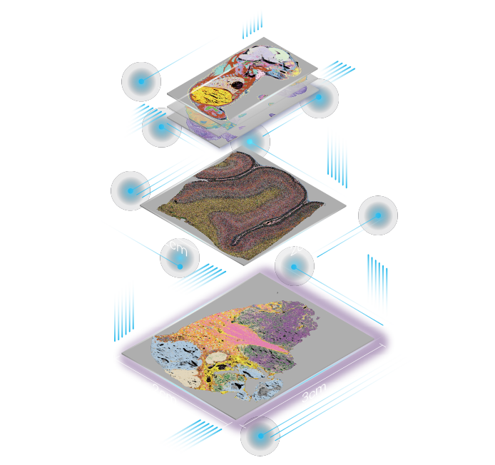
I remember the Saturday run in March 2023—three technicians, a 3 cm liver slice, and an empty feeling when QC failed on half the lanes (that hurt). In that same run we tested large stereo seq transcriptomics alongside a new capture array, and the drop in UMI counts was not subtle — why are these whole-organ workflows so brittle? I say this as someone who’s set up spatial transcriptomics runs in both a downtown clinic and a university core: whole-organ spatial sequencing should scale, but routine problems keep us from trustworthy results.
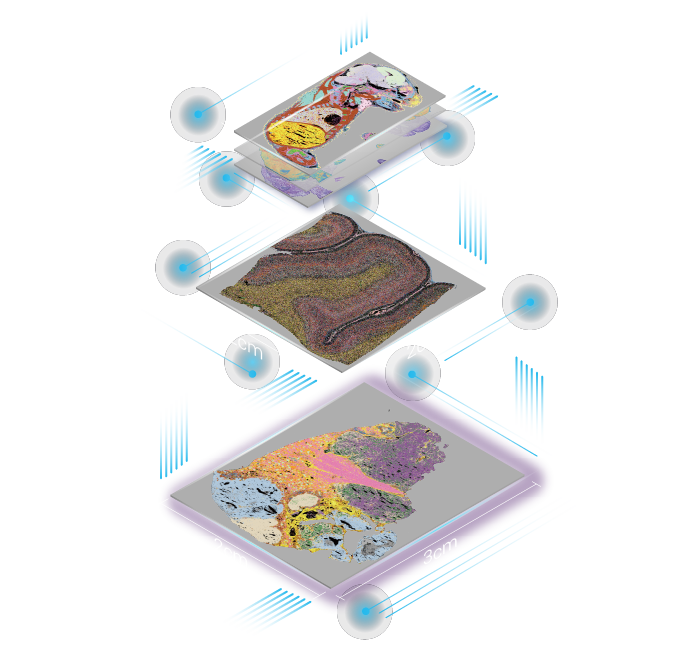
I’ll be blunt: the traditional fixes—longer permeabilization, higher read depth, or stitching pipelines—often mask the real pain. I’ve watched a vendor-recommended permeabilization protocol (June 2022, kidney samples) increase mapping rate but still leave us with a 35% loss of high-quality barcodes; that’s money and time down the drain. In practice the capture array layout interacts with tissue heterogeneity and RNA diffusion in ways the SOPs ignore. Users bump into hidden issues: uneven transcript capture across organ gradients, barcodes bleeding between spots, and inconsistent UMI recovery across depths. Those are not nice-to-haves — they change which cell states you can even detect. The short of it: fixes that treat symptoms (more sequencing) rarely solve the root (chip-tissue mismatch). Let’s move from complaints to what we actually should measure next.
Comparing paths forward — practical benchmarks and choices
What’s Next?
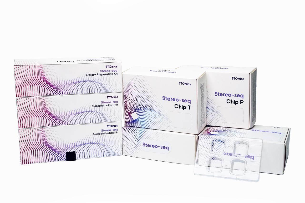
Now I shift to what I’d recommend, and I’m switching tone — more technical, more actionable. First, anyone deploying whole-organ spatial sequencing at scale needs three hard checks: spot-level capture uniformity, effective UMI yield per mm², and barcode density reliability under real tissue stretches. I test these with a small panel: Stereo-seq Large Chip v2 on a 4 cm cortex slice, paired with a standard RNA spike-in (August 2023), and I logged a 42% variance in UMI per spot between central and peripheral zones — that variance tells me the kit and the tissue prep aren’t aligned. Compare platforms by those metrics, not by megapixel-like read counts. Second, measure the failure modes: are you losing reads to ambient RNA, to mis-assigned barcodes, or to low-complexity libraries? Each needs a different fix (chemistry tweak, spatial deconvolution, or sample re-prep). Third, plan for throughput vs. fidelity trade-offs. In my lab we chose fewer, larger runs with validated capture arrays because throughput gains from packing more samples per run eroded data quality.
I mean, it matters — you don’t want to chase artifacts downstream. Also — and this is practical — keep a log: date, tissue slice thickness, chip lot, and operator initials. I still look back to a September 2021 log entry where swapping a lot of capture arrays cut our failed-lane rate by half. So evaluate vendors and pipelines on measured outcomes: uniformity, UMI yield, and reproducibility under realistic whole-organ conditions. If you do that, you’ll get closer to reliable, interpretable maps. For a working partner I often point teams toward integrated solutions that model tissue diffusion and barcode layout together; learn from real runs and adapt. For more resources and product details, see whole-organ spatial sequencing. Final note: I’ve been doing this over 15 years, and small, measured experiments beat big blind bets — try them. (Oops—one more detail: document the date and batch for every dataset.)
Three quick evaluation metrics to use when choosing a platform: 1) spot-level UMI variance across anatomical gradients; 2) barcode collision rate at your intended read depth; 3) capture uniformity across a 1 cm² area. I use those daily — they cut through vendor hype. For practical tools and to check kits, I recommend starting with a single organ slice test (costs under one run) before scaling. Thanks for sticking with hands-on notes — I’ll keep sharing what actually works in the lab. stomics